Diagnostic Methods
- Fourier-Transform Infrared Spectroscopy (FTIR)
- X-ray Photoelectron Spectroscopy (XPS)
- Mocon
- Scanning Electron Microscope (SEM)
- Defect diagnostics
- Multi Resonance Probe (MRP)/Plasma Absorption Probe (PAP)
- Energy Resolved Mass Spectrometry
- Phase Resolved Optical Emission Spectrscopy (PROES)
X-ray photoelectron spectroscopy (XPS)
At the Research Department Plasmas with Complex Interactions an X-ray photoelectron spectroscopy (XPS) device is available for surface analysis. The instrument is located at the Chair for Experimental Physics II in the NB building on the RUB campus. Measurements in the context of research progress on campus are supported by the XPS instrument by providing insights into bonding states at the surface of coatings or materials. For example, this provides insight into the progression of growth behavior of coatings. In addition, depth profiling provides the opportunity to look deeper into the layer. Research on surface diagnostics is currently being carried out in CRC 1316, which will provide information on the influence of atmospheric pressure plasmas on catalysts.
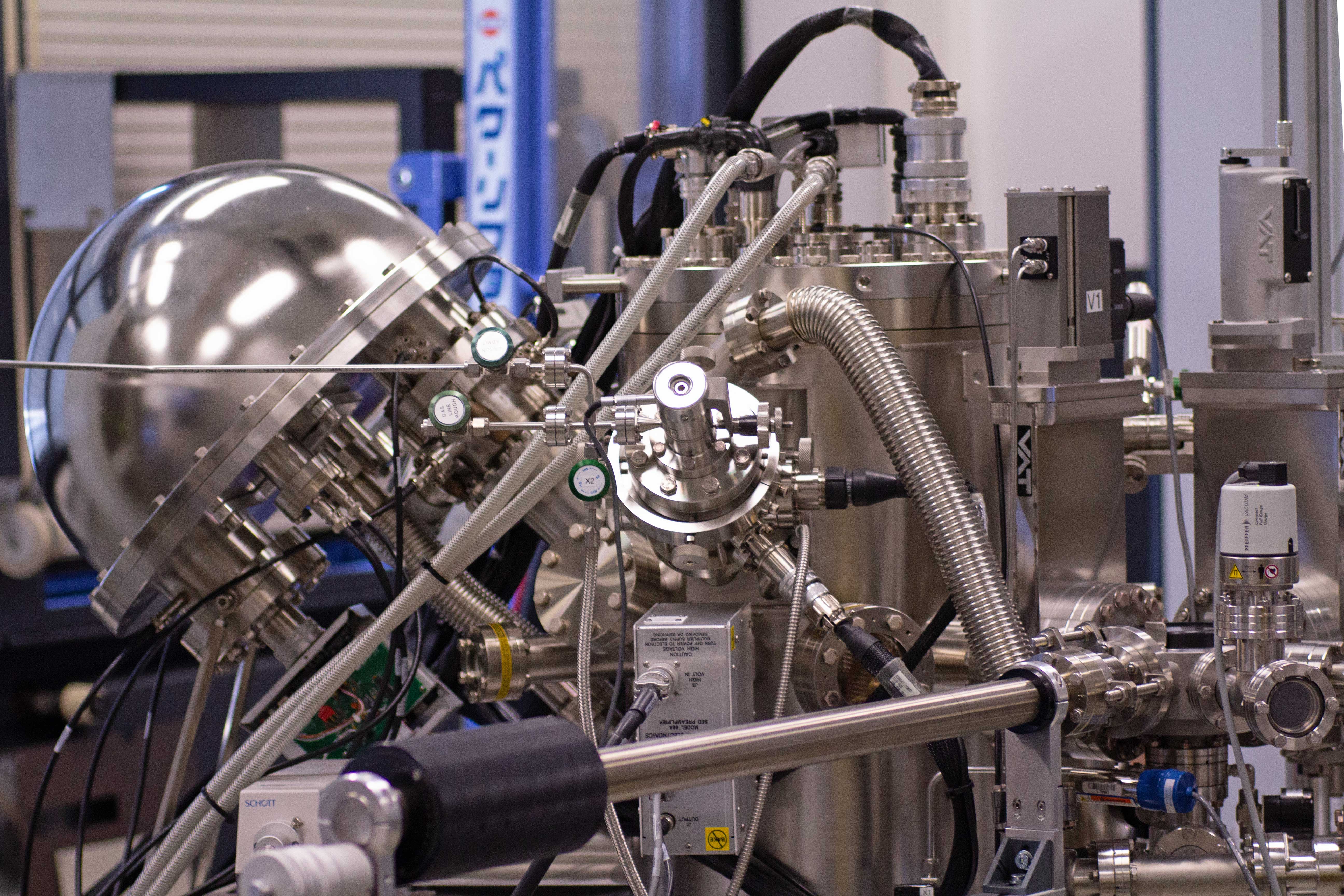
The operating principle of XPS is based on the extraction of electrons from a sample surface irradiated with X-ray photons. X-rays are generated when a beam of electrons with sufficient energy to promote transitions between atomic nuclear levels strikes an anode. The generated X-rays are directed to a monochromator where radiation with an energy of 1486.6 eV is selected. These X-ray photons are directed onto a sample. The interaction of the X-ray beam with the atoms of the sample leads to the excitation and extraction of electrons. The extracted electrons are collected in a detector where their kinetic energy is measured. Each element is characterized by the binding energies of the electrons in the different atomic nuclei levels. The only elements that are not detectable in XPS are hydrogen and helium. Chemical bonding between atoms results in a shift in the binding energy position, allowing chemical analysis of the measured samples. This technique is referred to as ESCA (Electron Spectroscopy for Chemical Analysis).
X-ray Photoelectron Spectroscopy is performed using a Versaprobe spectrometer from Physical Electronics (PHI 5000 VersaProbe). At an aluminium anode, Al K α radiation with an energy of hν = 1486.6 eV is produced. A resolution of 0.5 eV is achieved for survey spectra using a pass energy of 187.85 eV. A spectral resolution of 0.05 eV for a pass energy of 23.5 eV is usually applied for measurements of single peaks. Best resolution achieved is 0.025 eV. The measurement spot can be applied to diameters of 20 μm, 100 μm or 200 μm. Standard measurements are performed at a tilt angle of 45° between the sample and the detector. For angle resolved measurements, angles between 15° and 85° can be used. An ion gun with an argon surce is adapted so that a sputtering of the samples with energies between
20 V and 4 kV can be used.
Three kinds of sample holders are avaible, the small one is a circular 1 inch holder, the bigger one is a circular 2 inch holder. Finally, there is a angle resolved sample holder on which up to eight samples can be mounted. The circular sample holders have masks which can be installed to measure wafers or any other solid sample.
For measurement request, please write an eMail to
Fourier-Transform Infrared Spectrometer
IFS 66/5
The IFS 66/S is a Fourier Transform Infrared Spectrometer for investigation of the composition of gases, liquids and solids. The technique is based on the absorption of IR-radiation and changing the vibrational or rotational state.
This spectrometer has a resolution better than 0.25 cm-1 at a frequency range from 7500 cm-1 to 370 cm-1. The aperture range is from 0.25 up to 12.0 mm with a total of 16 different apertures. The source is a watercooled SiC glowbar for measurements in the NIR. The beamsplitter is a Ge multilayer coating on KBr. The velocity of the mirror allows a scan rate from 0.055 cm/sec up to 10 cm/sec and up to 20 spectra/sec at a resolution of 8 cm-1. The spectrometer affords the opportunity to measure with an internal detector in the FTIR itself or outside of the spectrometer with an external detector.
This spectrometer allows Step Scan measurements. These are helpful for time resolved spectroscopy of repetitive events with a time resolution between 10 ms up to 5 µs. Also polarization studies, photoluminescence spectroscopy, two dimensional IR spectroscopy and photo induced absorbance spectroscopy are possible.
IFS 55
The IFS 55 is a Fourier Transform Infrared Spectrometer for investigation of the composition of gases, liquids and solids. The technique is based on the absorption of IR-radiation and changing the vibrational or rotational state.
The spectrometer has a resolution of 0.5 cm-1 at a frequency range from 7500 cm-1 to 370 cm-1. It offers 16 scan velocities between 0.1 and 10.1 cm/s and a scan rate up to 20 spectra/sec at a resolution of 8 cm-1. The spectrometer affords the opportunity to measure with an internal detector in the FTIR itself or outside of the spectrometer with an external detector.
Energy Resolved Mass Spectrometer
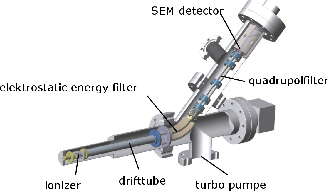
The Hiden EQP 300 HE is an energy resolved mass spectrometer which is used to measure time resolved ion energy distribution functions (IEDF) of high power pulsed magnetron sputtering (HiPIMS) discharges.
The mass spectrometer has a quadrupol filter which allows the detection of ion masses up to 300 atomic mass units (amu) with a precision of 0.01 amu. In its standard variation the energy filter, consisting of two parallel plates with a voltage apllied to, is capable of measuring the energy of ions in a range of -100 eV up to 100 eV in steps of 0.01 eV. The addition of the high energy component (HE) allows for measurements of ion energies from -1000 eV up to 1000 eV. An ionisator can be used for the detection of neutral particles.
A secondary electron multiplier is used as a detector to count ion rates. Attached to the analogue output of the detector is a transient recorder which is capable of counting events with a temporal resolution of 100 ns.
The mass spectrometer needs to be pumped separately from the vacuum chamber to which it is attached. The separate pumping reduces the amount of collisions that particles undergo in the mass spectrometer and, thus, increases the transmission of the particles.
Multi Resonance Probe/Plasma Absorption Probe
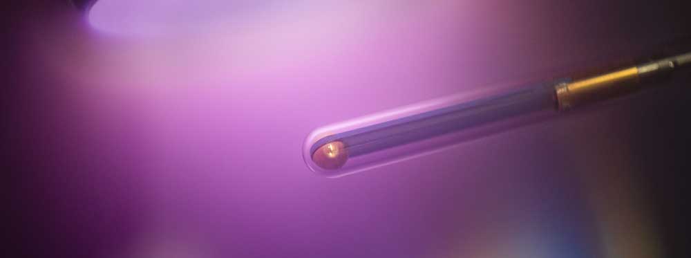
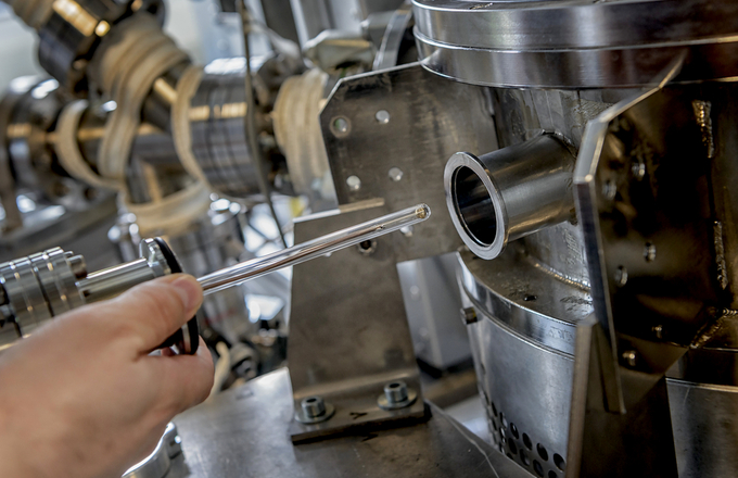
The head of the Multipole Resonance Probe (MRP) consists of two metallic hemispheres working as electrodes of a small antenna and separated by a dielectric layer. The head’s diameter is 6 mm and is enclosed by a dielectric tube (thickness of 1 mm) which is immersed in the plasma. This makes the probe insensitive against ceramic coatings which is needed in many applications. A self-developed electronic or, as an alternative, a network analyzer feeds an rf signal sweep to the antenna and displays the frequency dependence of the power absorption. This method is known as active plasma resonance spectroscopy (APRS). From the absorption spectrum the value of the electron density is calculated. In comparison to other APRS probes, the MRP shows a geometrical and electrical symmetry which significantly decreases the complexity of the model. The measurement range of the electron density is between 1013 m-3 and >1018 m-3. Time resolution lies in the range of sub ms, depending on the used electronic.
The plasma absorption probe (PAP) was invented as an economical and robust diagnostic device to determine the electron density distribution in technical plasmas. It consists of a small antenna enclosed by a dielectric tube which is immersed in the plasma. A network analyzer feeds a rf signal to the antenna and displays the frequency dependence of the power absorption. From the absorption spectrum the value of the electron density is calculated. The original evaluation formula was based on the dispersion relation of plasma surface waves propagating along an infinite dielectric cylinder. In this letter the authors present the analysis of a less idealized configuration. The calculated spectra are in good qualitative agreement with their experimental counterparts, but differ considerably from those predicted by the surface wave ansatz. An evaluation scheme which takes our findings into account will improve the performance of the PAP technique further.
The measurement range of the electron density is between 1014 m-3 and 1018 m-3. Time resolution lies in the range of sub ms.
PROES
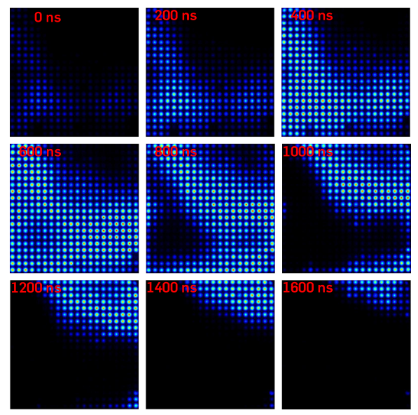
Phase resolved optical emission spectroscopy (PROES) is a technique that alows the investigation of the discharge emission according to the time, especially, phase of the applied excitation voltage. In addition, the combination of an ICCD-Chip and optical filter establishes the spatial resolution of the emission of a selected in wavelenghts.
Here, the camera (LaVison PicoStar HR16) works with a time resolution of about 100 ps. The ICCD-Chip has 512x512 pixels and enables a spatial resolution of few µm. The camera can detect emission in range between 350 nm and 750 nm.
Mocon
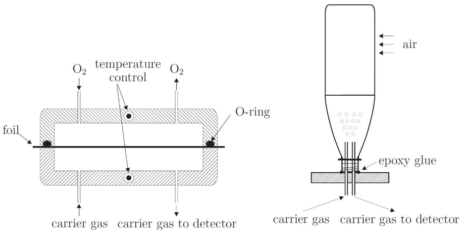
The MOCON OX-TRAN Model 2/61 is an oxygen permeation measurement system. Oxygen transmission through plastic foils or packages is analyzed in six independent cells by means of the carrier gas method. Each cell is sealed and consists of two parts which are separated by a plastic sample. One part is flushed with 100 % oxygen or ambient air. The other part is flushed with forming gas which serves as carrier gas. Oxygen which had permeated through the plastic, is transported to an electrochemical sensor responsible for the measurement. Oxygen Transmission Rate (OTR) can be obtained as a function of temperature in a range from 20°C to 65°C and as a function of relative humidity (RH) between 35 % to 100 %. Measurements are performed for plastic foils with a sample area of 10 cm² or plastic packages (like PET bottles).
